Nano-sized cluster bombs used in oncology
The illustration is taken from an interesting article on the color classification of chemotherapy drugs, which also I recommend read to visitors to this post.
Thus, chemotherapy is literally a weapon of mass destruction and initially acted at the cellular level as indiscriminately as chemical or nuclear weapons. Already in the second half of the 20th century, along with the development of immunotherapy, ways were sought to minimize the dose of radiation or drug so that the treatment would primarily target the tumor and not the entire body. Such research is carried out in the field of biotechnology and nanotechnology, and the principle of influencing the tumor varies.
Firstly, the tumor can be illuminated using special proteins, and then the active substance can be directed to the tumor.
Secondly, it is possible to enclose the active substance in a shell (nanocapsule), which will release the drug in the immediate vicinity of the tumor or inside it.
Thirdly, radiotherapy on a nanoscale is possible – supplying specially selected isotopes to the tumor, which have a short half-life, and decay products are easily eliminated from the body.
It is when using the second strategy that the drug carrier is often compared to a cluster bomb. Such a metaphor leads, for example, Israeli scientist Dan Peer from Tel Aviv University. We'll return to its development below, but first we'll discuss what was probably the first chemotherapy bomb of its kind, constructed in 2005 at the Massachusetts Institute of Technology. Its authors were Ram Sasisekharan (Professor of the Department of Biology at MIT) and his colleagues. Sasisekharan identified the most insurmountable dilemma of chemotherapy as the need to balance the toxicity of the drug's effects on healthy cells and overcome the drug resistance that mutating cancer cells acquire. Since Darwinian natural selection plays in cancer's favor here, the harmful effects of chemotherapy on the body are not reduced, and the tumor's susceptibility to the drug may be reduced.
As I wrote earlier in the article about thalidomide, the most important drug for inhibiting tumor growth is anti-angiogenesis, that is, counteracting the growth of blood vessels. It is this mechanism that largely explains the teratogenic effect of thalidomide on the human fetus. Thalidomide practically did not gain a foothold in this capacity, but the antiangiogenic payload for nanoparticles was considered an effective active substance, which, however, should not enter the bloodstream prematurely and damage healthy vessels.
The MIT team filled the outer membrane of an organic nanocapsule with an antiangiogenic drug and the inner cavity with a chemotherapy drug. These capsules had a diameter of only 200 nm, despite the fact that the diameter of a red blood cell varies from 7000 to 9000 nm. Such small capsules not only eluded the attention of the immune system, but also easily penetrated the tumor.
Inside the tumor, the outer membrane of the capsule quickly dissolves, and first the antiangiogenic drug works, and then the chemotherapy itself. The antiangiogenic drug acts so that the growth of blood vessels in the tumor stops, and the network of vessels inside it lasts longer, since it is through these vessels that the tumor has time to slowly become saturated with chemotherapy.
The first results on mouse models were encouraging, but later it turned out that such an effect actually leads to tumor shrinkage, but at the same time stimulates its metastasis and spread from a “dangerous place.” The overall result was rather unsatisfactory.
In 2010, Dan Peer and his co-authors succeeded in developing a nanosized carrier containing tiny granules of paclitaxel, which is used in the treatment of breast cancer, ovarian cancer, Kaposi's sarcoma and other neoplasms. The diameter of such a particle is 135 nm. When it comes into contact with a cancer cell, it goes off like a cluster bomb, pumping its payload into the cell.
From a scientific point of view, the most important thing in this preparation was to select the substance from which the nanoparticle shell is composed. This is a specific sugar recognized by receptors on the surface of the cancer cell. There are no such receptors on the surface of healthy cells, so the cluster bomb only affects malignant cells. In addition, having struck a cell, the nanoparticle is resorbed and then easily eliminated from the body, unlike traditional chemotherapy.
Actinium bombs
Already in 1995, David Sheinberg from the Memorial Center named after. Sloan-Kettering in New York tried to combine nanotechnology with radiotherapy. Sheinberg and his colleagues were able to seal an actinium-225 atom into a specially synthesized molecular cage, and then tested it on mice. The cell consisted of carbon and nitrogen atoms, the design looked like this:

The isotope actinium-225 is a by-product of the operation of nuclear reactors, so it is relatively easy to obtain:

Scheinberg's cell consisted of a dense ring lattice of carbon atoms, and inside it was a much larger actinium atom. The actinium atom has a negative charge, and the cell has a total positive charge, so the isotope is held in the cell as if by magnets. An antibody attaches to the cell from the outside, pulling it towards the tumor. The cell moves freely in the bloodstream until it finds a tumor. The half-life of actinium (10 days) is quite sufficient for a therapeutic effect, but all the decay products of actinium, especially francium, are also very radioactive (they emit alpha radiation), and the body cannot quickly remove them. However, actinium-225 is effective in treating leukemia, as well as reported in 2022, helps with inoperable prostate cancer. Due to the demand for the isotope, the State Research Center “Physics and Energy Institute” as part of the state corporation “Rosatom” began production of actinium-225, becoming the third scientific institution in the world where this isotope is specially synthesized.
Expansion of functions and illumination
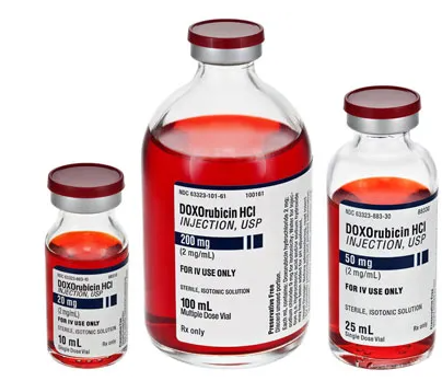
In 2016, a collaboration between China University of Science and Technology, Georgia Tech, and Emory University created the first two-level cluster bomb charged cisplatin, which has been used in chemotherapy since the early 1960s. Cisplatin is extremely toxic, causing pathologies of the kidneys and inner ear, but has been successfully used to treat many types of cancer, including neuroblastoma, retinoblastoma, tumors of the cervix, urethra, prostate and bladder. In this configuration, the cluster bomb “shell” has a diameter of 100 nm, but inside the tumor, even smaller cisplatin granules are released from it, which literally explode due to the difference in pH levels around and within the tumor. Small “bombs” break the DNA of a tumor cell, not just poisoning it, but directly preventing it from dividing.
In the same 2016, more subtle methods also began to be used in oncology, fundamentally similar to the use of the cluster bombs described above. A group of researchers from the Women's Hospital. Brigham in Boston, Massachusetts, has synthesized nanoparticles that begin emit light in the presence of dying cells. Such particles do not cause direct harm to the tumor, but provide quick feedback on whether the chemotherapy used is working and whether it primarily kills tumor cells or different types of cells throughout the body.
Conclusion
The research described above interested me, first of all, because of its bionic rather than nanotechnological nature. Cluster bombs for carrying anticancer drugs are fundamentally similar to red blood cells. An erythrocyte is a hollow cell filled with hemoglobin and, for greater capacity, devoid of any organelles. In the course of evolution, the red blood cell acquired just such a device, since free hemoglobin is toxic and, when entering the bloodstream, causes liver failure. These are the processes that develop when red blood cells are destroyed when the Rh factor does not match. The described cluster bombs (including those filled with sea anemone) have a shell of proteins or sugars, on which, like on a red blood cell, antigens and antibodies can be placed. Perhaps in the future, such technology will find application in genetic engineering for point and short-term genetic editing, as well as for the use of entire sets of borrowed genes in humans, so that these sets would be similar to bacterial plasmids.