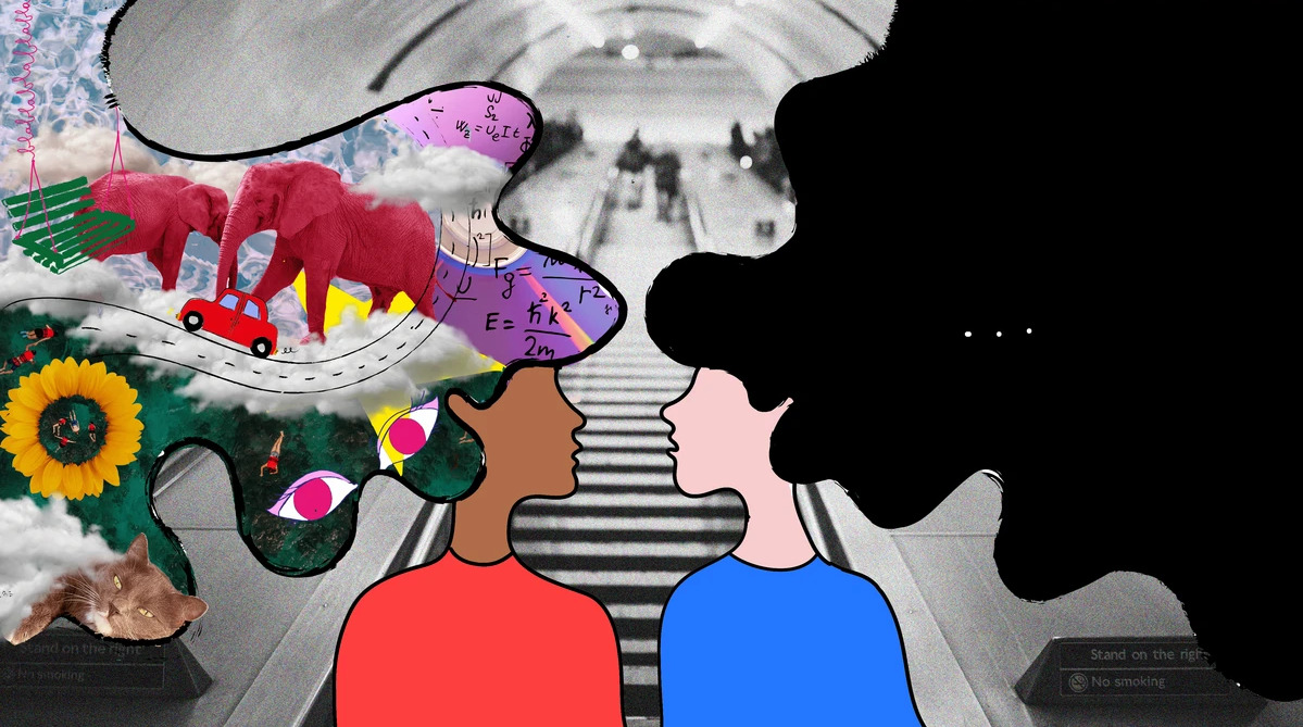
Long-term memory and aphantasia

Human memory is a very unusual mechanism, which is not yet fully understood. We may not remember what we did five minutes ago, but at the same time remember in detail the events of ten years ago. Autobiographical memories that concern us personally are formed in conjunction with visual images. The ability to create visual images of memories helps us re-experience certain events from the past, that is, remember them in more detail. However, there are people who are not able to create visual images. This deviation from the norm is called aphantasia. Scientists from the University Hospital Bonn (Germany) conducted a study in which they established which disorders in which areas of the brain lead to aphantasia. What experiments did scientists conduct, what results did they produce, and how can new data be used in practice? We will find answers to these questions in the scientists' report.
Basis of the study
Our unique and personal memories are stored in autobiographical memories (AM from
autobiographical memories
), ensuring stability and continuity of our “I”. For most of us, mentally traveling back in time and revisiting such unique personal events is associated with vivid, detail-rich mental images. These images during the re-experiencing of AM became a hallmark of autonoetic, episodic recovery of AM. However, to date it remains unclear to what extent episodic AM retrieval is dependent on visual imagery and what neural consequences the absence of mental imagery has on episodic AM replay. This knowledge gap exists because separating AM retrieval from mental imagery is challenging.
One way to solve this mystery is to study people with aphantasia. Recent research defines aphantasia as a neuropsychological condition in which people experience a marked reduction or complete absence of voluntary sensory imagination. Individuals with aphantasia thus provide a unique opportunity to study the consequences of episodic AM retrieval in the absence of voluntary imagination.
From a neural perspective, the hippocampus is considered a central brain structure supporting detail-rich episodic AM retrieval in the healthy brain. In fact, hippocampal activity correlates with the vividness of AM memories, and patients with hippocampal damage show marked impairments in detailed episodic AM retrieval. In addition, neuroimaging studies show that the hippocampus is almost always coactivated with a broader set of brain regions, including the ventromedial prefrontal cortex (vmPFC from ventromedial prefrontal cortex), lateral and medial parietal cortex, and visual-perceptual cortex. Interestingly, especially during the AM retrieval stage, when people actively retrieve episodic details from a specific AM, the hippocampus shows strong functional connectivity with the visuoperceptual cortex. This indicates the crucial role of this connection for the reproduction of visuoperceptual details in AM.
However, there are few studies examining the neural correlates of aphantasia. From the few available, it was concluded that there is potential hyperactivity of the visual-perceptual cortex in aphantasia. The current hypothesis states that due to this hyperactivity, small signals occurring during mental imagery may not be detected. If this is true, the impairments in episodic AM production observed in aphantasia may be due to altered connectivity between the hippocampus and visuoperceptual cortex. The authors of the work we are considering today decided to test the assertion that the AM deficit observed in aphantasia is due to changes in the hippocampus, visual-perceptual cortex and their functional connection.
Preparing for the study
The study involved 31 people who had no previous history of mental or neurological diseases. Of these, 15 had congenital aphantasia, and 16 acted as a control group.
Aphantasia is usually assessed using the Visual Vividness Questionnaire (VVIQ from Vividness of Visual Imagery Questionnaire), i.e. a subjective self-assessment questionnaire that measures how vivid mental scenes a person can create. For example, people are asked to visualize a sunset in as much detail as possible and rate their mental scene on a 5-point Likert scale (ranging from “no image at all, you just ‘know’ what you think about the object to “perfectly clear and vivid image”). The highest score on the VVIQ is 80, which indicates the ability to visualize mental images as vividly as if the event were happening right here and now. The minimum score is 16, which indicates that the person was unable to create any mental image at all. Aphantasia is at the lower end of the imagery spectrum and is usually diagnosed with a VVIQ score of 16 to 32.
Given that the VVIQ questionnaire is highly dependent on subjective self-assessment, the scientists decided to also conduct a binocular competition test to increase the objectivity of assessing the ability to create mental images. After presenting either a red-horizontal or a blue-vertical Gabor pattern, participants were presented with a red-horizontal Gabor pattern for one eye and a blue-vertical Gabor pattern for the other eye. Subsequently, participants were asked to indicate which type of Gabor pattern they predominantly observed. Typically, successful mental imagery leads subjects to select the Gabor pattern they just visualized. This selection bias can be translated into a priming score that reflects the strength of the visual images. Imitation stimuli consisting of only red-horizontal or blue-vertical Gabor patterns were presented on 12.5% of trials to allow for detection of decision bias.
Next, the study participants underwent an autobiographical interview, during which they were asked to remember any event from a certain period of their life: before 11 years old, 11-17 years old, 18-35 years old, 35-55 years old and a year ago. Participants under 35 years old, instead of the period 35-55 years old, recalled the events of the previous year. Memories of the first four periods were considered distant, whereas memories of the previous year were considered recent.
The interview was structured in such a way that each memory consisted of three parts: a free-form story, generalized details, and specific details of the event. During the first stage, a person spoke in any form about an event from a particular period of his life. During the second stage, the person had to name as many details of that event as possible. And during the third, answer specific questions from the interviewer (time, place, emotions/thoughts at that moment). Participants were then asked to rate their memories in terms of their ability to visualize the event on a 6-point Likert scale (from “not at all” to “very well”).
To assess the details of memories, they were divided into two categories: internal details and external details. Internal details included: internal events (incidents, weather conditions, people present, actions), place (country, city, building, part of the premises), time (year, month, day, time of day), details of perception (visual, auditory, gustatory, tactile, olfactory, body position) and emotions/thoughts (emotional state, thoughts). Subcategories of external details were semantic details (factual or general knowledge), external events (other specific events in time and place but different from the main event), repetition (repetition of identical information without prompting), and other details (metacognitive utterances, editorial utterances). In addition, following a standard procedure, each memory was assigned an episodic richness rating on a 7-point Likert scale (ranging from “not at all” to “perfect”). Confidence in one’s memories was also assessed on a 7-point Likert scale (from “not at all” to “perfect”).
Finally, participants were asked to answer three questions:
- How difficult is it for you to recall autobiographical memories?
- How difficult is it for you to navigate in space?
- How difficult is it for you to use your imagination?
Participants answered each question on a Likert scale from 1 (“very easy”) to 6 (“very difficult”).
Next, an fMRI test was conducted, during which the subjects were asked to either remember an event in detail or give an answer to a simple mathematical problem. During recall tests, cue words such as “party” were presented on the screen for 12 seconds, and participants were asked to recall a personal event associated with the cue word that was specific in time and place (for example, a party for their 20th anniversary). When the participant was ready to give an answer, he pressed a button. After their response, participants also rated the vividness of that memory.
During the problem test, simple addition or subtraction problems, such as 47 + 19, were presented on the screen for 12 seconds. Here, participants had to press a button after the problem was solved. Next, they had to add 3 to the previously obtained solution, for example, (47 + 19) + 3 +… + 3. After this, the participants also rated the complexity of the text.
The MRI protocol consisted of anatomical scans, resting-state scans, and fMRI sessions conducted while performing tasks from the AM tests.
Research results

Image #1
People with aphantasia scored significantly lower on the VVIQ than controls. In addition, the test and control groups differed in their starting scores on the binocular competition task. While the control group was attuned to their own mental images on 61.3% of trials, people in the test group were tuned in only 52.6% of the time. In fact, the performance of controls was significantly different from chance, whereas the performance of people with aphantasia was not. Moreover, VVIQ scores were positively correlated with performance on the binocular competition task. These findings support the fact that the strength of visual imagery was reduced in people with aphantasia.
In an autobiographical interview, people with aphantasia reported fewer memory details than controls. They were also more likely to recall recent events rather than distant ones. Internal details were mentioned more often than external ones. More importantly, a two-way interaction was found between memory detail type and group. This indicates that people with aphantasia reported significantly fewer internal memory details, but not significantly fewer external memory details, compared to controls (1b). There was no two-way interaction between memory period and group, and no three-way interaction between memory period, group, and memory detail type.
Based on the interaction effect between group and memory detail type, the researchers compared specific categories of internal and external memory details between groups. In terms of internal details, compared with controls, people with aphantasia reported fewer internal details, fewer emotional details, fewer perceptual details, and fewer details regarding time and place (1c). On the other hand, no significant differences were found in external details, including accompanying events, semantic details, repetitions, etc. In terms of assessments, people with aphantasia reported less vivid memories and less confidence (1a) in the accuracy and detail of their memories, and also poorly assessed their ability to visualize memories.
When answering the three questions above, there were pronounced differences between the groups of subjects. Although people with aphantasia reported that they typically had great difficulty recalling autobiographical memories and using their imagination in everyday life, they did not report difficulties with spatial orientation.
The scientists found dramatic differences in the intensity of the response between the groups. While controls reported on 86% of trials that their AM memory was vivid, people with aphantasia said so on only 20% of trials. Moreover, people with aphantasia responded more slowly than controls when asked whether the memories they retrieved were vivid or not. In contrast, during the math task trials, there were no significant differences between groups in either easy/difficult response or reaction time.
In addition, people with aphantasia and controls spent similar amounts of time retrieving memories in AM trials. Moreover, the two groups did not differ in their speed of solving math problems. People with aphantasia spent 3.87 seconds solving a math problem, while the control group spent 3.90 seconds.

Image #2
One of the main objectives of this study was to examine AM-related hippocampal activity in subjects with aphantasia and controls (image #2). Extracted fMRI signals illustrate dramatic differences between subject groups in AM-related hippocampal activation. People with aphantasia showed decreased activation in the bilateral hippocampus, including the left anterior, left posterior, right anterior, and right posterior hippocampus. There were no lateralization effects or differences in activation patterns along the anteroposterior axis between groups. These data indicate that AM behavioral deficits associated with aphantasia are reflected neuronally by decreased bilateral hippocampal activation.

Image #3
The researchers also sought to examine whole-brain activation during autobiographical memory retrieval in both groups (3a And 3b). The tables below show the coordinates of AM and MA activation for people with aphantasia (Table 1) and for the control group (Table 2).

Table No. 1

Table No. 2
Overall, both groups showed greater activation in all regions typically associated with AM, including the bilateral hippocampus, ventromedial prefrontal cortex, and medial/lateral parietal area, during autobiographical memory retrieval. When examining differences between groups, people with aphantasia showed greater activation in bilateral visual-perceptual areas (maximum in the lingual gyrus) in the occipital lobe than controls (3c, 3d, 3e). In contrast, the control group showed greater activation in the right hippocampus. Additional correlation analyzes showed that participants with higher activation in the visual-perceptual cortex had less activation in the hippocampus. This suggests a trade-off between increased activation of the visual-perceptual cortex and decreased activation of the hippocampus.

Image #4
Whole-brain analysis supported the hypothesis that the main difference between people with aphantasia and controls was the interaction between the visual-perceptual cortex and the hippocampus. To test this interaction, the researchers examined the functional connectivity between hippocampal and visual-perceptual cortex peak differences during autobiographical memory retrieval (snapshots above). Scientists found differences in functional connections between the right hippocampus and the left visual-perceptual cortex. Interestingly, while people with aphantasia show little to no functional connectivity between these two regions, controls showed strong negative connectivity.

Image #5
Finally, the scientists decided to take a closer look at the resting-state connectivity between the identified regions of interest in the right posterior hippocampus and left/right visual-perceptual cortex. There were no differences in the activity of these areas between the groups of subjects. Interestingly, in the control group, a positive correlation was found between resting-state connectivity of the right hippocampus and visual cortex and interview imaging scores. On the other hand, in people with aphantasia, a negative correlation was found between resting-state connectivity of the right hippocampus and visual cortex and interview imaging scores.
Overall, fMRI results indicate that the impairment in autobiographical memory retrieval associated with aphantasia is reflected by functional changes in the hippocampus and visuoperceptual cortex, as well as interactions between them.
For a more detailed understanding of the nuances of the study, I recommend taking a look at scientists' report.
Epilogue
In the work we reviewed today, scientists tried to establish a connection between the activity of certain parts of the brain and ability. A person visualizes his memories. Scientists paid special attention to people with aphantasia, that is, a lack of visual imagination (the ability to mentally create images).
The study found that changes in two important brain regions, the hippocampus and the occipital lobe, and their interactions play a role in the impairment of personal memory retrieval in aphantasia. Previous neurobiological studies have shown that the hippocampus, which acts as a brain buffer during memory formation, supports both autobiographical memory and visual imagination. However, the relationship between these cognitive functions has not been previously established.
The study included people with and without aphantasia. During the experiments, both groups had to remember a memory from the past, describe it in as much detail as possible, and then evaluate their own story. Observations have shown that people with aphantasia have a more difficult time recalling memories. Not only do they provide fewer details, but their stories are less vivid and their confidence in their memory declines. This suggests that the ability to remember autobiographical events is closely related to imagination.
As subjects tried to describe the memory in as much detail as possible, their brain activity was recorded using fMRI. The scans showed that the hippocampus, which plays an important role in recalling vivid, detailed autobiographical memories, was less activated in people with aphantasia. Differences were also identified in the interaction between the hippocampus and the visual cortex, which is responsible for processing and integrating visual information in the brain.
The main conclusion of this work, as its authors note, is to confirm in practice the fact that people with a reduced ability to mentally visualize certain images have a much more difficult time remembering events associated with them. In the future, scientists plan to determine whether people who are blind from birth can create detailed autobiographical memories. An even more important question, according to scientists, is whether this skill can be developed through training. If this is true, it may be possible to help people suffering from memory disorders such as Alzheimer's disease by training visual imagery instead of conventional memory training.
A little advertising
Thank you for staying with us. Do you like our articles? Want to see more interesting materials? Support us by placing an order or recommending to friends,
cloud VPS for developers from $4.99
,
a unique analogue of entry-level servers that we invented for you:The whole truth about VPS (KVM) E5-2697 v3 (6 Cores) 10GB DDR4 480GB SSD 1Gbps from $19 or how to properly share a server?
(options available with RAID1 and RAID10, up to 24 cores and up to 40GB DDR4).
Dell R730xd is 2 times cheaper in the Maincubes Tier IV data center in Amsterdam? Only here 2 x Intel TetraDeca-Core Xeon 2x E5-2697v3 2.6GHz 14C 64GB DDR4 4x960GB SSD 1Gbps 100 TV from $199 in the Netherlands! Dell R420 – 2x E5-2430 2.2Ghz 6C 128GB DDR3 2x960GB SSD 1Gbps 100TB – from $99! Read about How to build a corporate infrastructure. class using Dell R730xd E5-2650 v4 servers costing 9,000 euros for pennies?


