It’s hard to imagine the life of a modern person without a computer. Remember the days when processors were single core? One of these processors – Intel Pentium 4 – began to be produced in 2000. Of course, such processors have long been out of use and now they can only be found among those who like to collect such things. So we at Smart Engines went through the employees and found several copies. What for? The answer is simple. We really wanted to look at the processor from the inside. We made a tomography of the processor using a domestic tomograph, and received reconstruction and 3D visualization using an Elbrus processor. This is how the tomography of Intel Pentium 4 on Elbrus turned out. In the article we will talk about expectations, our actions and the results obtained.
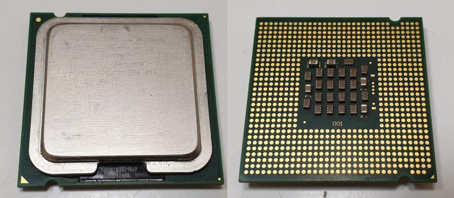
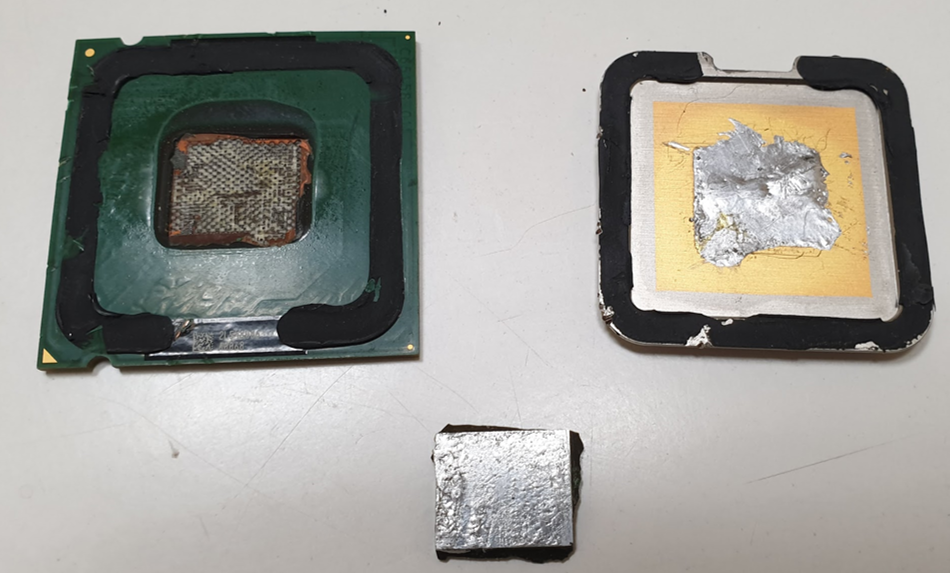
The heart of the processor is a silicon chip hidden under a metal cover that strongly absorbs X-rays and must be removed in order to “illuminate” the crystal. Using a gas torch and a screwdriver, the cover was removed, and the crystal itself was torn off from the substrate.
The processor before the start of the study, top and bottom views
Crystal (silver rectangle) prepared for research
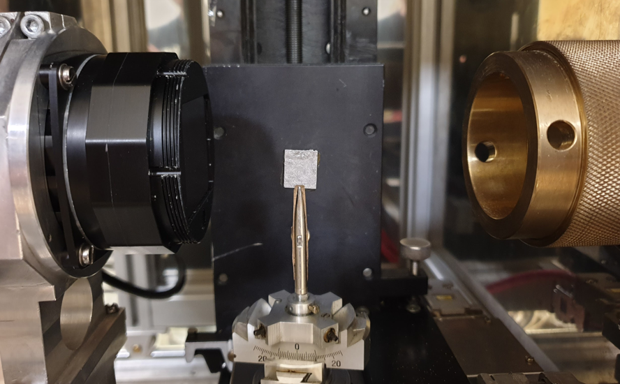
Crystal in the tomograph
We carried out tomographic measurements on a laboratory tomograph designed and assembled at the Federal Research Center “Crystallography and Photonics” of the Russian Academy of Sciences (Federal Research Center KF RAS) in the laboratory of reflectometry and small-angle scattering (we talked about it here).
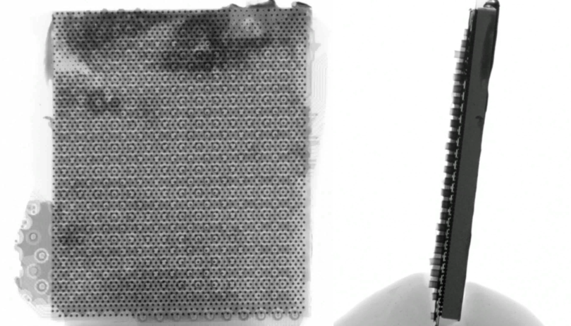
Because metal layers remained above and below the crystal, then radiation with an energy of 40 keV was chosen for tomography, on the one hand it penetrates the metal, and on the other hand, silicon is not completely transparent for it. 800 images of the crystal were obtained with a resolution of 9 μm.
Examples of X-ray images of a processor crystal
The processor seemed to be a difficult object to study, because in one direction it is elongated and strongly absorbs, and if it is rotated 90 degrees, it will be thin and almost transparent to X-rays.
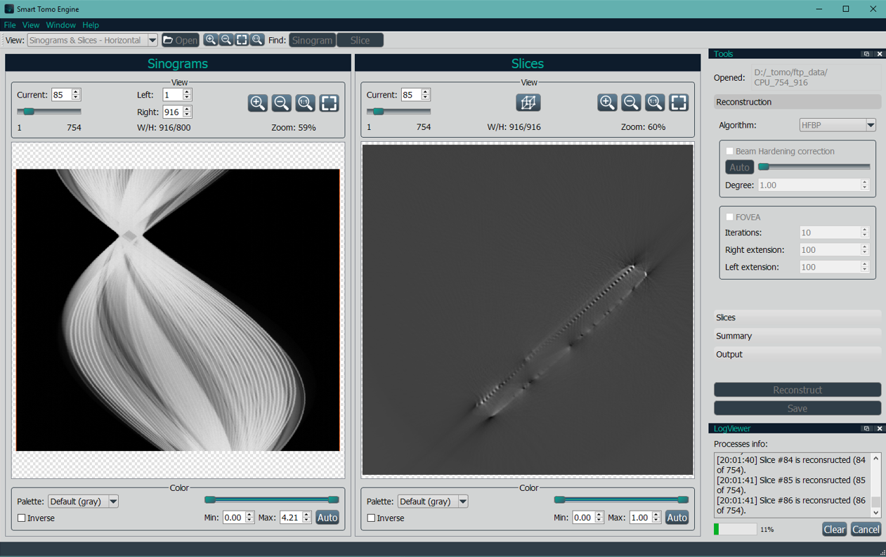
Further, the registered set of images had to be processed. The reconstruction was carried out using the Smart Tomo Engine program developed by us on the Elbrus-4C machine. For reconstruction, we used the HFBP algorithm, we talked about it here.
At the stage of data preprocessing, the data size was reduced by half, so we got 800 frames of size 754 by 916. We reconstructed 754 layers, the size of one layer is 916×916. View of pre-processed sinograms and reconstruction of one layer.
Screenshot of Smart Tomo Engine
Here’s a reconstruction we got:
Looking at the reconstructions obtained, we were convinced that our algorithms for tomographic reconstruction allow us to avoid “metal like” artifacts when examining objects containing both strongly and weakly absorbing regions (metal and silicon).
As a conclusion
Remember how earlier, in childhood, we often took apart various electro-mechanical toys (cars, robots and moon rovers) to see how they work there, and, possibly, find some familiar parts (light bulbs, motors). We studied the designs of these “complex” devices, analyzing the interposition of the “macro” elements, completely excluding the details that were incomprehensible at that time in the form of capacitors and resistors.
So it is now. Only instead of lunar rovers – processors, and instead of a screwdriver – tomographs. Although we understood in advance that with a tomograph with a resolution of 10-15 microns, we would not see the structure of transistors (after all, in the production of a Pentium 4 crystal, 90 nm technology was used), the desire to look inside an interesting object, study its structure and understand its constituent parts (even not all) is something that does not disappear from the inquisitive brain of the researcher.