An alternative method of transpedicular fixation or as a caliper can replace an entire x-ray machine
Currently, spinal surgery has ceased to be something exclusive and is performed in almost all departments of neurosurgery and in many trauma departments of hospitals. The term “spinal instability” has long gone beyond the narrow circle of spinal surgeons. And although this concept is sometimes interpreted too broadly, instability of the spinal segment, as a pathological phenomenon, exists. This article will discuss one interesting method that simplifies spinal surgery.
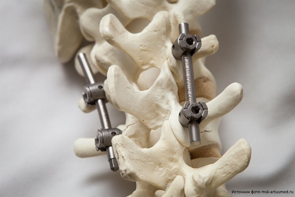
Transpedicular fixation (TPF) is used when, due to physical, dystrophic and any other changes, the spine is not able to fully perform its supporting functions. It can be fractures, deformations and displacements of the vertebrae, cancer with the defeat of its individual sections, etc. Before the operation, an individual selection of the supporting structure is carried out taking into account the patient's condition and the presence of pathologies. During the surgical intervention, the surgeon fixes the wire frame on the segments of the spine with special screws that are inserted into the leg of the vertebra (pedicle), which, by the way, gave the name to this method.
Reliable fixation of the vertebrae and spinal column with the help of high-strength titanium screws can significantly reduce the patient's rehabilitation period, speed up the process of restoration of his disability.
TPF methods and their problems
The transpedicular systems today are quite diverse, but they all have only one goal: the most correct way to insert screws into the vertebral body. Each installed screw should in no case go beyond the boundaries of the cortical layer of the vertebra, otherwise it can damage the adjacent nerve and vascular formations, as well as permanently damage the vertebra without the possibility of its further recovery. The decisive in this sense is the choice of introduction points and deviations of the trajectory in the axial and sagittal planes.
For example, consider the most common ways to create spinal fusion.
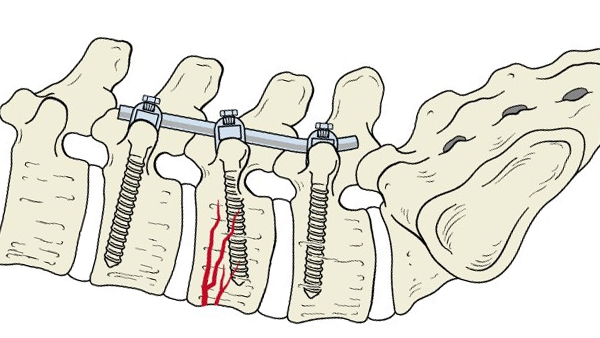
• Freehand technique. The method of installing screws, based on the determination of points and angles of insertion by anatomical landmarks in real time. For the operation, the surgeon relies solely on his personal experience and skills, so the quality of the intervention depends entirely on the level of professionalism of the doctor. In addition, the duration of such an operation is difficult to predict, since the anatomy of each spine is very variable. In each case, it is necessary to select individual angles and points of introduction, which increases the time of the operation, and therefore the length of time the patient is under anesthesia, which can not be called useful.
• X-ray assisted. A method of performing an operation in which surgery is divided into stages, an X-ray examination of the operated area of the spine is performed between them in order to timely monitor the path of insertion of the screws and prevent their improper installation. Obviously, during the operation it is necessary to perform several x-rays, which increases the radiation dose to the patient and staff. The need to interrupt surgery to take an image also poses a risk of sterility disruption.
• Modern technologies. The use of various robotic and navigation tools, intraoperative control systems, can significantly increase the accuracy of screw installation, reduce the duration of the operation. The main disadvantage of such techniques is their traditionally high cost.
The presence of the voiced difficulties, the need to increase the accuracy of the installation of transpedicular screws, reduce the cost and duration of the operation, are forcing to develop new methods for the effective creation of spinal fusion.
Alternative method
In order to achieve high accuracy of the installation of transpedicular screws using simpler methods, the neurosurgeon doctor of the neurosurgery department of the Clinical Hospital No. 1 of the Presidential Administration of the Russian Federation Kalyuzhny Vasily Gennadievich proposed an alternative method of surgery, which involves preoperative planning based on processing of studies obtained using computer tomography (CT). The essence of this method is to find the optimal trajectory of the introduction of screws using computer simulation of the operation. To process images, special software is required, which we agreed to develop specifically for this task. The benefit was a lot of experience in such development for medicine.

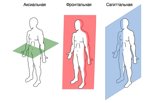
Today, DICOM is the generally accepted standard for storing, transmitting and visualizing medical research. Our program works with such DICOM data. Images obtained as a result of CT examination are usually located in the axial plane parallel to each other. However, for the purpose of preoperative control, it is also important for the doctor to see the general picture of the features of the spine in other planes. To solve this problem, multiplanar reconstruction (MPR) is used – a special processing of the source data, during which it is possible to obtain images in the sagittal, frontal or any arbitrary plane.
During the planning of the operation, the doctor selects the problematic vertebra and selects a section in the axial plane. An additional slice is also displayed in the frontal plane and three sagittal. In addition, for a more detailed assessment, correction of points and trajectory of screw insertion, a set of nine sections is made in the horizontal plane with an interval of 2 mm vertically. This technique helps to fully take into account the anatomical features of a particular vertebra in order to exclude surgical errors during the operation.
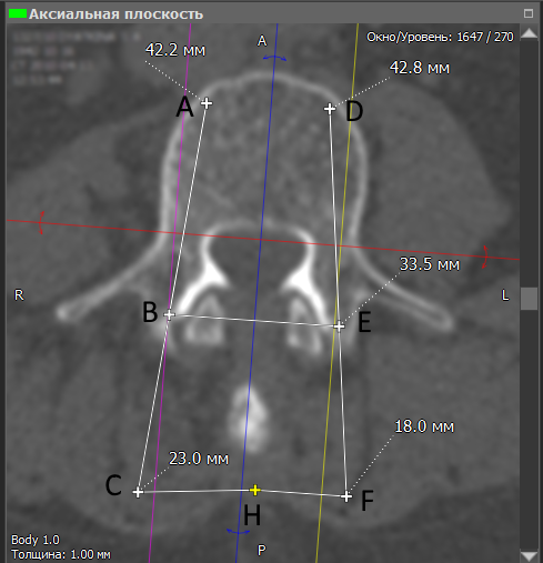
Next, the user draws on the problem vertebra lines of the trajectory of the introduction of screws AC and DF. Points B and E are points of insertion of screws into the vertebra, and the resulting segments A-B and D-E determine their future position. The projection of the screws is displayed in all planes, which allows you to additionally adjust the paths of their insertion, which in one plane may be optimal, but in others it may turn out to be undesirable or even dangerous.
Having determined the position of both screws, the doctor marks the point H on the spinous process in the picture, which is needed to determine points C and F. The program automatically determines the lengths of the desired segments, which are displayed in millimeters. Knowing the points of insertion of the screws (B and E), as well as the required distances from the spinous process (segments C-H and H-F), the doctor during the operation can determine the paths along which it is necessary to drill the pin channel to install the screws in the right position.
Having performed correction taking into account the three-dimensionality of the vertebra during the multiplanar reconstruction, the doctor receives marking cards for the upcoming operation, which can be printed on a conventional printer. Thus, the surgeon gets directly into the operating room with a detailed plan of what and how to do.
During the operation, the subcutaneous tissue is dissected in the area of the problem area and the vertebra is accessed. Next, marking occurs: in accordance with the picture, using a vernier caliper on both sides of the spinous process, points are determined that, together with the known screw insertion points, form trajectories along which the position of the tool for forming the pin channel is controlled. Next, drilling and direct insertion of transpedicular screws takes place.

Measuring the distance between the pin channel forming tool and the apex of the spinous process to control the angle of entry in the axial plane

To clarify the quality of the operation and create a picture of the rehabilitation prognosis, all patients undergo mandatory postoperative CT monitoring.
Advantages of the Alternative TPF Method
Compared with existing methods of restoring spinal functions using transpedicular screws, the proposed method shows several important advantages at once.
• High accuracy of screw installation – about 97.1% of correct insertion, which is comparable with navigation methods for carrying out such operations.
• Reducing the duration of the operation due to preliminary planning and layout using effective computer technology.
• Significant reduction in radiation exposure to the patient and staff due to the lack of the need for continuous x-ray control, characteristic of other methods.
• The possibility of additional detection of anatomical and pathological formations due to preoperative CT control.
Thus, the proposed method of transpedicular fixation of the spine allows relatively simple means to perform the operation to restore the functions of the musculoskeletal system with good accuracy, comparable with similar indicators of more complex methods. In 2017, patent RU2620355C1 – A method of installing screws for transpedicular stabilization of the spine, was obtained.
Despite the development of technology in the field of medicine, you should not rely on the fact that any problem can be solved on the operating table. It is better to worry about your own health in advance and not bring the matter to a critical state. And then it is not necessary, for example, to strengthen the spine beyond what was provided for by nature.